Microscopy is the technical field of using microscopes to view samples and objects that cannot be seen with the unaided eye (objects that are not within the resolution range of the normal eye). There are three well-known branches of microscopy: optical, electron, and scanning probe microscopy.
Microscopes are devices that are used to magnify small objects. They come in a wide range of shapes and sizes, and use many types of illumination sources (light, electrons, ions, x-rays and even mechanical probes) and signals to produce an image. A microscope can be as simple as a hand-held magnifying glass or as complex as a multi-million-dollar research instrument.
Microscopists explore the relationships between structures and properties for a very wide variety of materials ranging from soft to very hard, from inanimate materials to living organisms, in order to better understand their behavior.
Optical and electron microscopy involve the diffraction, reflection, or refraction of electromagnetic radiation/electron beams interacting with the specimen, and the subsequent collection of this scattered radiation or another signal in order to create an image. This process may be carried out by wide-field irradiation of the sample (for example standard light microscopy and transmission electron microscopy) or by scanning of a fine beam over the sample (for example confocal laser scanning microscopy and scanning electron microscopy). Scanning probe microscopy involves the interaction of a scanning probe with the surface of the object of interest. The development of microscopy revolutionized biology and remains an essential technique in the life and physical sciences.

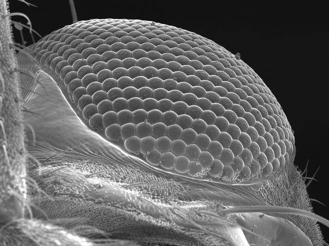
SEM Micrograph of the compound eye of a house fly
Contact Us
Tel & Address Info
Tel: 012 420 2075
Email: admin@microscopy.co.za
Address:
University of Pretoria,
Cnr Lynwood and Roper Street, Hatfield
Pretoria, 0028, South Africa
Operation Hours:
Monday - Friday
07h 00 - 15h 30


